Table of Contents
Uncover the secrets of milialar, or milk spots—exploring causes, types, prevention, and treatments. This guide empowers all age groups for healthier, clearer skin.
Milia, often referred to as milium cysts or milk spots, are small, benign lumps or cysts that can appear on various parts of the skin, with a preference for the face. While generally harmless, they can be a cosmetic concern for many individuals. In this comprehensive guide, we will delve into the intricate details of milialar, exploring its types, causes, prevention strategies, and treatment options. Whether you’re a concerned parent of a newborn or an adult seeking flawless skin, this guide aims to empower you with the knowledge needed to understand, prevent, and treat milia effectively.
What is Milialar?
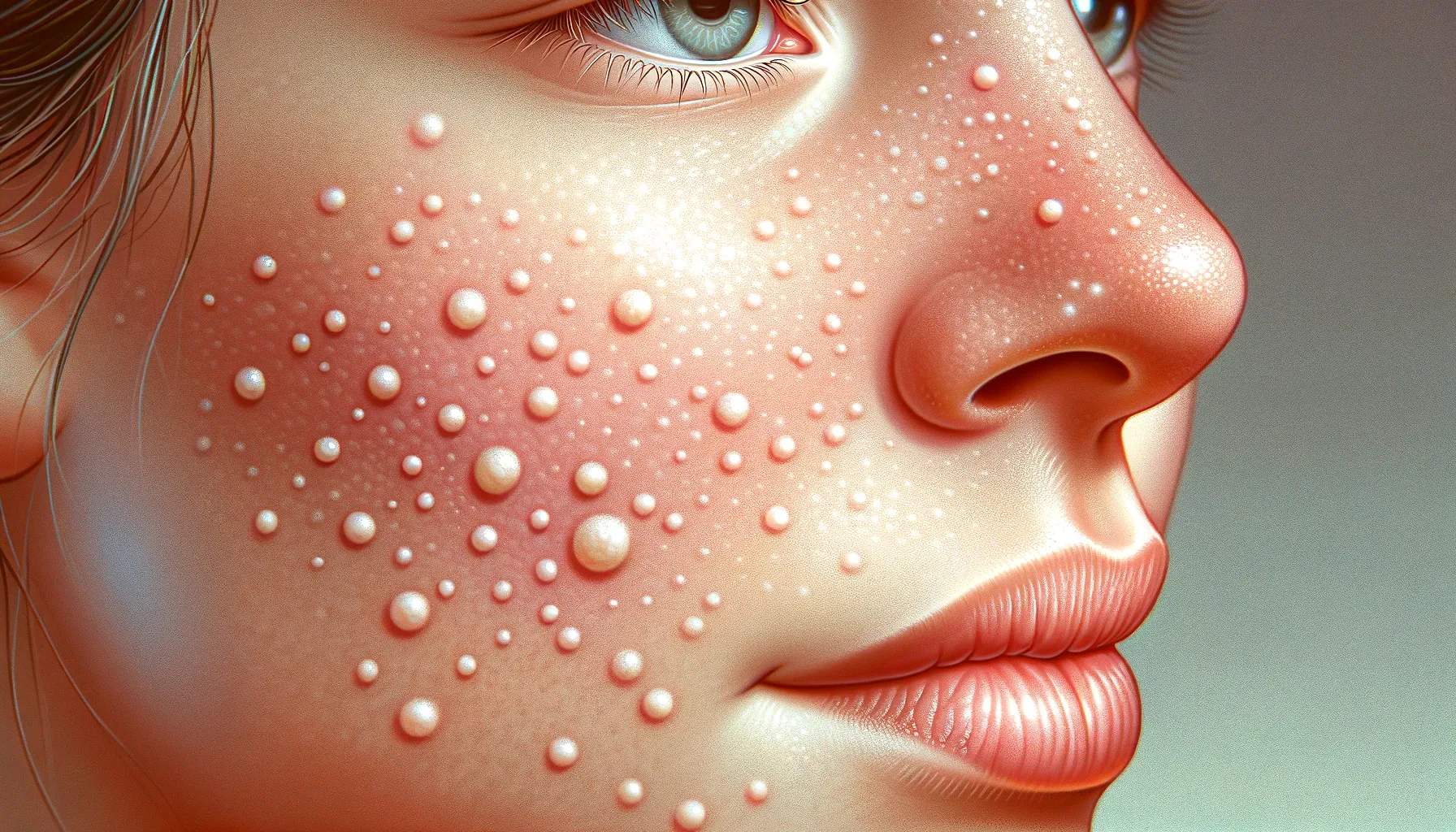
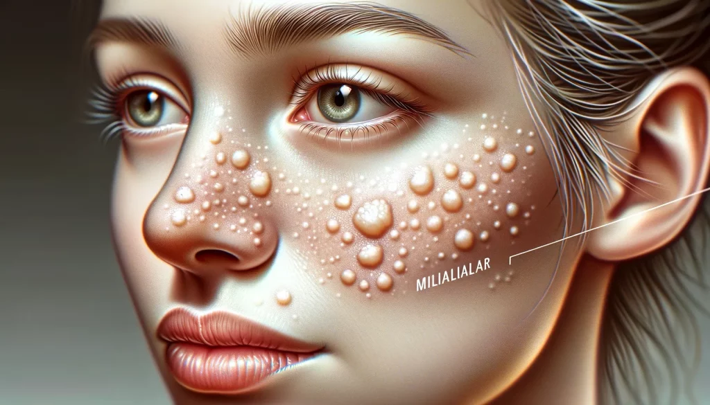
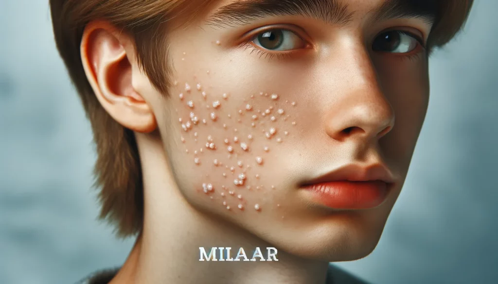
Milia are small, dome-shaped bumps that often manifest as whitish-yellow, pearly cysts on the skin’s surface. Ranging in size from 1-2 millimeters, these cysts resemble tiny pearls embedded under the skin. The most common locations are around the eyes, on the eyelids, cheeks, nose, and forehead. Milialar develops when dead skin cells become trapped beneath the skin’s surface, forming keratin-filled cysts. While typically harmless, they can be a cosmetic concern, prompting the exploration of prevention and treatment options. Read also Winona Health Physical Therapy.
Types of Milialar
1.1 Primary Milialar
Primary milialar occurs when sweat ducts are blocked by dead skin cells, commonly seen in newborns due to immature sweat ducts. These small, white to yellow cysts are often found on the cheekbones, nose, and around the eyes.
1.2 Secondary Milialar
Secondary milialar arises from skin damage or wounds, such as burns or blisters. It can also appear after certain skin treatments in adults and may require varied treatment approaches depending on the underlying cause.
1.3 Neonatal Milia
Neonatal milia is a common condition affecting newborns soon after birth. These harmless cysts typically resolve on their own within a few weeks.
1.4 Milia en Plaque
This rare type of milia is characterized by a cluster covering an irritated, elevated area of skin.
Causes of Milialar:
Understanding the factors contributing to milialar development is essential for effective prevention. Several common causes include:
2.1 Excessive Sun Exposure:
Prolonged exposure to the sun can damage facial skin, increasing the risk of milia formation.
2.2 Skincare Choices:
The use of oily or heavy skincare products can clog pores, leading to milia. Opting for non-comedogenic products is crucial for prevention.
2.3 Skin Trauma:
Any form of skin trauma, such as burns or blisters, may result in secondary milia.
2.4 Genetics:
Genetic predisposition plays a role in certain individuals being more prone to acquiring milia.
2.5 Sweating and Humidity
Excessive sweating and high humidity levels can obstruct sweat ducts, contributing to milia development.
Development Process
Understanding the specific process of milialar development sheds light on its origins:
3.1 Skin Renewal:
As part of its renewal process, the skin naturally sheds dead cells. However, sometimes, these cells don’t shed properly.
3.2 Trapped Keratin:
The trapped cells form keratin, accumulating beneath the skin’s surface.
3.3 Formation of Cysts:
This accumulation leads to the formation of tiny cysts, resulting in the characteristic appearance of milia.
Expert Insight: There are several subtypes of milialar, with primary milialar arising spontaneously due to keratin entrapment being most common around the eyelids. Secondary milialar can arise from trauma, burns, blistering, or ophthalmic conditions.
Signs and Symptoms of Milialar:
Recognizing the signs and symptoms of milia is crucial for early identification and potential treatment. Milialar is generally easy to recognize with the following characteristics:
4.1 Appearance:
- Small, pearly white bumps on the eyelids or around the eyes.
- Dome-shaped, smooth bumps resembling pearls under the skin.
- Whitish-yellow or yellowish-white in color.
- May appear singly or in clusters.
4.2 Sensation:
- Typically painless and doesn’t cause itching or irritation.
- Can remain unchanged for weeks to months or disappear on their own.
- Sometimes may secrete a waxy, cheese-like discharge if ruptured.
Tip: If uncertain about any skin condition or experiencing concerns, consulting a dermatologist for a proper diagnosis and guidance is advisable.
5. Preventive Measures:
While it may not be possible to prevent milialar entirely, adopting the following preventive measures can be helpful in reducing the risk of development:
5.1 Skincare Practices:
- Use oil-free, non-comedogenic moisturizers and makeup.
- Avoid heavy, greasy creams and cosmetics near the eyes.
- Cleanse gently and exfoliate the skin regularly to unclog pores.
- Shave carefully using proper technique to avoid injuring the skin.
5.2 Sun Protection:
- Wear sunscreen daily and limit unprotected sun exposure.
5.3 Hydration:
- Keep your skin well-hydrated to prevent excess dryness.
5.4 Makeup Removal:
- Remove makeup thoroughly before bedtime and discard old makeup.
5.5 Tailored Treatments:
- Treat any underlying skin conditions like eczema.
- If prone to milia, consider avoiding intensive facials or chemical peels which may worsen them.
Treatment Options for Milialar
While milialar often resolves spontaneously, various treatment options are available for persistent cases:
6.1 Prescription Retinoid Creams:
- Creams containing tretinoin, adapalene, or tazarotene can help dry out and slough off milia.
6.2 Microdermabrasion:
- This technique uses fine crystals to gently exfoliate the outer skin layers and stimulate healing.
6.3 Chemical Peels:
- Applying a mild glycolic or salicylic acid solution helps soften and remove the lesions.
6.4 Dermatological Extraction:
- A dermatologist can open the cyst with a sterile needle and squeeze out the contents.
6.5 Cryotherapy:
- Freezing the bumps with liquid nitrogen to eliminate the lesions.
6.6 Laser Ablation:
- Using laser energy to destroy the cysts.
6.7 Surgical Removal:
- In some cases, surgical removal by cutting open and draining the milia may be considered.
Important Note: This information is provided for educational purposes, and it is recommended to seek professional advice from a dermatologist before pursuing any treatment.
Final Words
Final Words, milialar, the small pearl-like cysts that can appear on the skin, are generally harmless but can be a cosmetic concern. While common in newborns, adults can also develop them, often as a result of skin damage. Milialar comes in different types, and their development is influenced by various factors, including genetics, sun exposure, skin trauma, medical conditions, medications, and the use of heavy creams and makeup.
This comprehensive guide has provided you with a detailed understanding of milia, its types, development, signs and symptoms, preventive measures, and treatment options. If you have any concerns about your skin or milialar, consult a dermatologist for professional guidance.
People also ask
Q: Can milia be prevented entirely?
A: While complete prevention may not be possible, adopting proper skincare practices and avoiding pore-clogging cosmetics can reduce the risk.
Q: Do milia always require treatment?
A: In many cases, milia resolve on their own. However, persistent or bothersome cases may benefit from various treatment options.
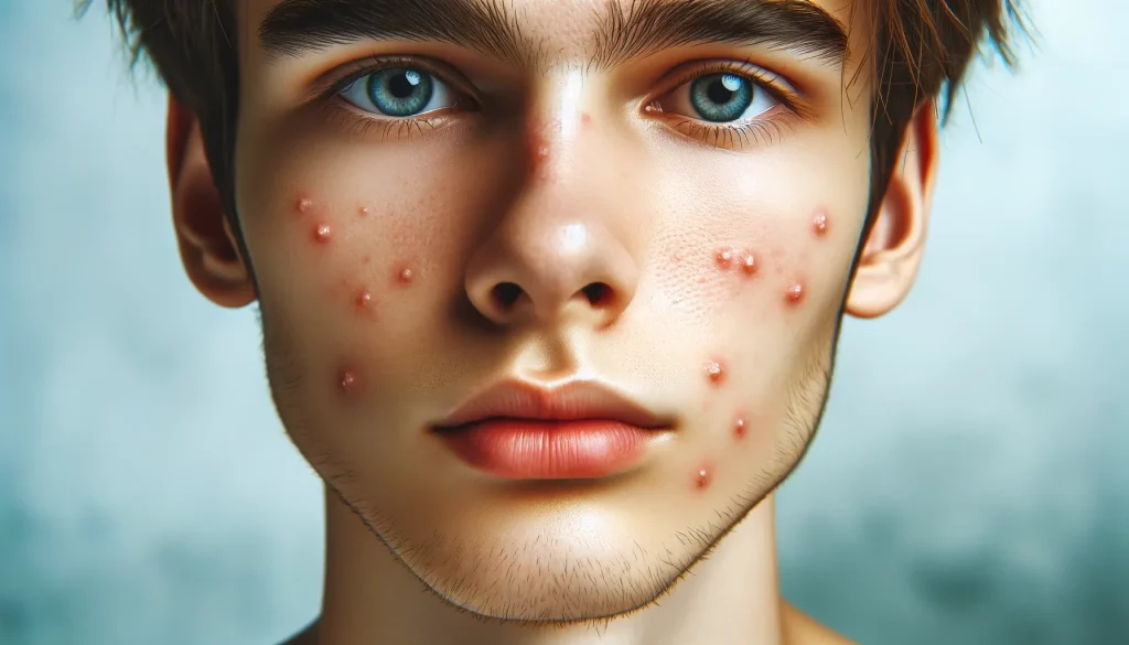
Q: Are milia related to acne or other skin conditions?
A: No, milia are distinct from acne. They are small cysts filled with keratin and are not associated with common skin conditions like acne.

Liam Stephens is a dynamic and skilled blogger, recognized for his ability to identify trends and create compelling content. As the founder of Remi-Portrait.com, Liam has become a reliable source of information across various fields such as food, technology, health, travel, business, lifestyle, and current events. He specializes in delivering up-to-date technology news and insights, catering to the diverse community that surrounds Remi-Portrait.com. His proficiency and engaging writing style have earned him a dedicated audience, solidifying his reputation in the digital sphere.